This section summarises the different treatment options available for intraocular and extraocular tumours. After leaving clinic, you may have forgot to ask a question about the treatment advised. This is very common when being faced with a diagnosis of eye cancer. We hope this section will help answer your questions. Please see the different treatment options below.
Intraocular Tumour Treatments
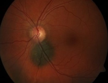
Below is the list of treatments available to us for tumours that grow inside the eye. The most common tumour that grows inside the eye is melanoma. The most common treatment for this is plaque radiotherapy. If the tumour is too big for this treatment we may consider proton beam radiotherapy. We aim to select the best treatment suited to each patient as an individual. Unfortunately this sometimes means having to remove the eye (enucleation). Please see the different treatment options below.
Plaque Radiotherapy
Plaque radiotherapy is a form of internal radiotherapy. A radioactive piece of metal known as a plaque is attached to the sclera (white part of the eye) next to the tumour. This is around the size of a 5 pence coin (please see picture above). This is done in the operating theatre and is left in the eye between 3 and 7 days before being removed. Patients usually stay in ward 1C for this treatment. The tumour starts to shrink around 4-6 months after the plaque is removed.
The effects can last for several years. Although being an effective treatment, the radiation can sometimes damage other parts of the eye. This may cause cataract, retinal detachment, nerve damage, or macular oedema (swelling of the back of the eye). New blood vessels may grow after treatment; these can sometimes block the drainage angle in the eye causing glaucoma. If we are unable to control this, we may have to consider removing the eye. Although there are risks of plaque radiotherapy, this treatment can stop growth of the tumour in around 80% of cases.
Proton Beam Radiotherapy
This is where radiation with charged particles called protons are targeted at the tumour from outside the eye. This treatment is used if the tumour is too large or located too far back in the eye for plaque radiotherapy to work. As this treatment is highly specialised, the Douglas Cyclotron Unit, Clatterbridge, Liverpool is the only centre in the United Kingdom where the treatment is given.
Please follow the link to the patient journey through proton beam therapy.
If proton beam radiotherapy is the best option for you, we will organise transport to Clatterbridge and accommodation for you in near the Hospital (please see the team photo of the Clatterbridge team and the hospital above). This involves two separate visits to the Clatterbridge. At the first visit a treatment mask is made and fitted- this helps the team target the radiotherapy at the correct part of the eye. At the second visit, one to two weeks later, the treatment is given. You will likely travel down Sunday evening and return Friday later that week. Final measurements are made on the Monday and then the radiation is given over the remaining four days during (Tuesday to Friday).
Each treatment session takes around 20 minutes and is pain free. Before going down to Liverpool, however, we have to perform a small operation on the affected eye in Gartnavel General Hosptial, Glasgow. This is where we stitch tantalum markers (small metal discs smaller than a paper clip- please see photo above) to the sclera (white part of the eye) next to the tumour. We usually do this under General Anaesthetic. This helps the team target the radiation treatment more effectively when you go down to Clatterbridge. Proton beam radiotherapy takes a little longer to work than plaque radiotherapy.
We usually wait six months to see if the tumour starts to decrease in size. Although effective at treating tumours, the radiation can also damage normal parts of the eye and tissues around the eye when given. This may cause loss of eyelashes, loss of pigmentation of the eyelids, and inflammation of the conjunctiva causing a watery eye. Sometimes small blood vessels can grow at the back of the eye and into the drainage angle at the front of the eye. This can cause the pressure to build up in the eye and cause glaucoma. If we are unable to control the pressure and the eye becomes very uncomfortable, unfortunately we may have to consider removing the eye.
External Beam Radiotherapy
This is where radiation from a machine is targeted at cancer cells from outside the body. We find this treatment useful in patients that have cancer else where in the body that has spread to the back of the eye. On the other hand, if there has been a large tumour inside the eye that has grown behind the eye, we may choose to use radiotherapy after removing the eye. This treatment is spit into fractions.
This means the treatment is divided into smaller doses and spread out over a few days. This gives healthy cells in the body a chance to recover between treatments. If we are using radiation to treat the eyes or lymph nodes around the head and neck, then a special mask is usually made for patients. This mask is worn during treatment and helps aim the radiation at only the cancer cells. Although this helps prevent damage to healthy cells around the tumour, treatment may still cause loss of eyelashes, loss of pigmentation of the eyelids, and inflammation of the conjunctiva causing a watery eye.
Laser Transpupillary Thermotherapy

Transpupillary Thermotherapy (TTT) uses an infrared laser beam to heat the tumour up and kill the cancer cells. This technique is useful if there is uncertainty if the suspicious area is a melanoma or a naevus, or if the choroidal melanoma is small and radiotherapy is inappropriate due to poor health.
This treatment is sometimes combined with plaque radiotherapy as it reduces swelling and leakage from the blood vessels. After TTT the tumour gradually shrinks down if successful. Repeat treatment may be required at 6 months.
Possible complications from this treatment include retinal detachment, blockage of blood vessels, growth of new blood vessels, iris burns and cataract. Unfortunately tumour recurrence after treatment is common. This is more likely to occur if the melanoma is thick, close to the optic nerve, or non-pigmented.
Photo Dynamic Therapy
A light sensitive dye is injected into the blood stream. As the dye travels through the blood vessels to the back of the eye through the blood vessels, a special light is shined into the eye. This activates the dye and causes the abnormal blood vessels to close, shrink, and stop leaking.
This is useful in patients with a naevus or melanoma that is leaking fluid and building up at the back of the eye (macular oedema). Although this is not the main treatment for choroidal melanoma, we may find it useful in some cases where radiotherapy treatment is not possible.
Removal of Eye: Enucleation
Enucleation is the medical term for removing the eye. This is recommended when the tumour is too large for other treatments or has started to invade behind the eye. Removing the eye along with the tumour is sometimes preferred if there is a lot of pain and discomfort in the eye. This can be due to high pressure inside the eye caused by blockage of the drainage angle by tumour or new blood vessels grown after radiotherapy.
The idea of having your eye removed is scary. With current technology, however, we can get excellent cosmetic results with uniquely designed and fitted artificial eye implants (please click below to see photos). Our artifical eye clinic is run in Gartnavel General Hospital, Glasgow. Alternatively, some patients may prefer to just wear an eye patch after the tumour is removed.
After removing the eye, although this gets rid of the tumour growing in the eye, unfortunately this does not prevent the tumour growing else where in the body later in life. The most likely place for melanoma to regrow is the liver. For this reason we may decide to organise a liver ultrasound scan every year at your local hospital to screen for cancer growth.
Artificial Eyes
After the eye is removed, a temporary cosmetic shell is fitted in the operating theatre (see first picture above). We choose a colour to match the patient’s other eye. Although not a perfect colour match, this temporary shell will remain in place until the final artificial eye is made.
Once the eye socket has healed, the temporary cosmetic shell is removed and the final artificial eye implant can be fitted. This is done in the prosthetic eye department in Gartnavel General Hospital, Glasgow. The team will take photos of your normal eye. The artificial implant is then painted in fine detail to match the photo of your healthy eye. Please see the two other photos above (note both the healthy eyes have been dilated in clinic to examine the back of the eyes so the pupil sizes do not look symmetrical).
Removal of Eye and contents of Eye Socket- Exenteration
Exenteration means removing the eye with the tumour and the soft tissue around the eye. This treatment is required if the cancer has spread behind or around the eye. Sometimes the eyelids or part of the bone around the eye have to be removed if the tumour has invaded here. If this is the case, we will likely perform the operation at the Queen Elizabeth University Hospital with the help of our Oral and Maxilofacial Surgeon colleagues. Sometimes this treatment is combined with radiotherapy or chemotherapy. Our medical oncology team will help us choose the best treatment for you.
Excellent cosmetic results can be achieved after the tumour is removed. This involves further reconstructive surgery and being fitted with an artificial eye. Some patients, however, may prefer just to wear an eye patch or leave things as they are instead of having further surgery. After the cancer is removed we can plan treatment that suits your needs.
Extraocular Tumour Treatments
Please see below the list of treatment options for tumours that grow outside the eye. Every case is different. In clinic we will discuss the best treatment or combination of treatments for you.
Surgical Excision of Eyelid Tumours
This can be performed under local or general anaesthetic and as a day case. After tumour removal as much normal tissue is left behind to help keep the eyelid looking as normal as possible. The tumour is sent to the pathology laboratory to confirm the type of tumour and if it has all been removed. Eyelid tumours (mainly basal cell carcinomas) may be removed by a dermatologist (skin specialist). This is where a small part of the skin is removed then inspected under a microscope straight away. If cancer cells are still visible, then more tissue is removed and inspected again. This is repeated until there are no more cancer cells seen under the microscope. This helps remove as little normal tissue as possible mean while ensuring all the cancer is removed.
After the tumour is removed the eyelid is reconstructed to get the eyelid looking and functioning as normal as possible. If the tumour removed was small then this can usually be done on the same day. If the tumour removed was large and a lot of the eyelid had to be removed then reconstruction may be done on a different day. Skin or tissue can be taken from the other eyelid or from other parts of the body to re-form the eyelid. We commonly use the skin in front of the ear or from the inner surface of the upper arm. Sometimes we use tissue from the inner surface of the cheek- this heals very well after surgery. As there are many options, we aim to choose the best treatment option for you.
Freezing treatment: Cryotherapy
This freezes the tumour helping destroy the cancer cells. This can be used in combination with surgical excision, or on its own if surgery is not an option. Cryotherapy can is usually done the operating theatre under local or general anaesthetic. Treatment lasts several minutes. Although helping prevent tumour growth, no samples are sent to the lab so confirmation of tumour death is not always possible. If this is the case, we will monitor you carefully in the clinic. Sometimes we have to repeat this treatment more than once.
Radiotherapy
This treatment involves targeting the cancer with high energy radiation beams. This kills the cancer cells and stops them multiplying. This is used if surgery is not possible, for example, if the patient is too unwell or desperately does not want surgery. It may, however, be the preferred treatment of choice; for example, in lymphoma. This treatment is performed as an out-patient. This treatment is spit into fractions. This means the treatment is divided into smaller doses and spread out over a few days. This gives healthy cells in the body a chance to recover between treatments. If we are using radiation to treat the eyes or lymph nodes around the head and neck, then a special mask is usually made for patients. This mask is worn during treatment and helps aim the radiation at only the cancer cells. Although this helps prevent damage to healthy cells around the tumour, treatment may still cause loss of eyelashes, loss of pigmentation of the eyelids, and inflammation of the conjunctiva causing a watery eye.
Chemotherapy
Mitomycin C (MMC)
This is a chemotherapy drug that is applied to the surface of the tumour in theatre. MMC works by sticking the cancer cells’ DNA (the cell’s genetic code) together, stopping the tumour or cancer cells from growing.
This is applied to the surface of the cancer cells and therefore side effects of chemotherapy such as nausea, vomiting or hair loss are not experienced.
If MMC eye drops are being used, however, this can irritate the eye. We may give lubricant or steroid drops to treat this.
5-fluorouracil (5-FU)
This treatment, also called imiquimod, is a chemotherapy drug which is applied to the surface of skin tumours. Sometimes we use this to treat eyelid tumours. This drug causes the body’s immune system to produce a chemical called interferon. This attacks and kills cancer cells. It may irritate the skin when applied, this means the treatment is working. It is applied 3 – 5 nights a week and treatment can last up to 6 weeks.
